THE BRAIN
T
|
he Human brain is by far the most complex structure to
be ever build. Its complexity has dazzled majority of the people since last
century and is still the topic of research.
The adult brain weighs about 1.3 – 1.5 kg and is
approximately 2% of or total body weight, with Surface area of approximately
1120 cc in males and 1130 cc in females. In spite of its small size it contains
millions and billions of neurons that carry out daily functions of the body and
may be create someone like Einstein whose brain is still preserved for research
purposes.
The basic functional unit of brain is called as a
neuron. It is a specialised type of cell that has the capacity to conduct and
transfer electrical impulses, but once damaged can take years to regenerate.
There are various types of neurons and are classified
according to –
A) The
number of dendrites/processes
B) The
length of axons
According to number of processes they are
1. Unipolar
2. Bipolar
3. Pseudounipolar
4. Multipolar
According to length of axons they are
1. Golgi
type 1(Long Axon)
2. Golgi
type 2(Short Axon)

Fig 1.0 Types of neuron, based on number of processes.
Note: The bipolar neuron is not to be associated with
the Bipolar condition. Bipolar condition or disorder is psychological and is
characterised by fluctuant changes in the mood of person from happy to being
depressed. And has certain criteria where you can assess yourself (will be
discussed later).
The brain is made up of three components
1. Forebrain
2. Mid
brain
3. Hindbrain
All of these parts are evolved from a particular
embryological tissue which after development leaves certain parts behind and
are called as the ventricles and aqueducts
of the brain and the spine
(discussed further).
The Forebrain is made up of the following structures –
1. Cerebrum
2. Basal
ganglia
3. Hippocampus
4. Thalamus
5. Hypothalamus
6. Pineal
Body
7. Infundibulum
The Midbrain consists of
1. Crus
cerebri
2. Tegmentum
3. Tectum
The Hindbrain consists of
1. Pons
2. Cerebellum
3. Medulla
oblongata

Fig 1.1 Brain and its parts
The brain has 3 predominant functions
1. Motor
2. Sensory
3. Association
The Brain is located inside the skull, because of its
shape, the skull is modified inside to accommodate the lobes and the structures.

Fig 1.2 Superior view of Skull (Source: Pearson
Education, Inc, publishing as Pearson Benjamin cummings).
The brain is also divided according to lobes it has –
1. Frontal
lobe
2. Temporal
lobe
3. Parietal
lobe
4. Occipital
lobe
There is a hidden lobe called insula which is present
in between the temporal lobe and parietal lobe and is situated underneath the
parietal lobe.
The brain is thrown into folds that increase the
surface area. The grooves are called sulci (singular: Sulcus) and folds are
called gyri (singular: Gyrus).
The two halves of the brain are separated by the
Longitudinal fissure and falx cerebri, creating a right and left cerebral
hemisphere and the cerebral hemispheres are separated from the cerebellar
hemispheres by Tentorium cerebelli (a fold of dura matter).

Fig 1.3 Sulci, gyri and longitudinal fissure

Fig 1.4 Showing tentorium cerebelli and Falx cerebri.
The Brain is covered with 3 layers of tissue called as
the Meninges. It is this layer that is inflamed and thus causing meningitis.
The three meninges are –
1. Dura
Matter
2. Arachnoid
Matter
3. Pia
Matter
The Dura Matter being the outer most, lies below the
skull bone and carries the sinuses of the brain (venous drainage pathways). The
dura is made of 2 layers where the outermost layer being the supportive and
attaches to the skull and the inner one covering the brain and the other two
meninges.

Fig 1.5 Showing dura, tentorium and sinuses in cadaver
(posterior view).
The Arachnoid matter runs along the whole surface of
the brain covering it entirely but not going deep; where as the Pia matter runs
deep down into each and every sulcus and carries the blood supply.

Fig 1.6 Meninges simplified (source
Neuroscientifically challenged – YouTube).
As mentioned earlier, the Brain has sulci and gyri,
there are a few which are predominant and are used as landmarks to study all
the important parts of the brain.
The main sulcus used as a landmark is Central Sulcus
which separates the frontal lobe and the parietal lobe.
Another sulcus used as landmark is the lateral sulcus
which separates the Frontal and parietal lobes from the temporal lobe,
The other sulcus present are the parieto-occipital
sulcus, occipito-temporal sulcus, superior and inferior sulci, superior and
inferior temporal sulci, etc.
The image below shows the two main sulci used as
landmark.

Fig 1.7 Sulci of brain (blue colour indicating the
main sulci) [source – Sobotta’s textbook and atlas of human anatomy]
The main gyri used as landmarks and are of prime
importance because they have functional importance to study are –
1. Precentral
gyrus
2. Postcentral
gyrus
3. Superior
temporal gyrus
The precentral gyrus is located just in front of the
central sulcus and contains the motor functions of the body. The postcentral
gyrus is located just behind the central sulcus and contains the sensory
functions of the body. Superior temporal gyrus is located just below the
lateral sulcus and has the auditory functions of the body.
The figure shows the gyri mentioned above.

Fig 1.8 Gyri of brain.
The area that connects the two halves of the brain is
called as corpus callosum.
It is a band of Neurons that connect the right and
left half of the brain. It contains –
1. Rostrum
2. Genu
3. Body
4. Tail/Splenium
Each of them connect a specific part of the brain, and
if this corpus callosum was cut, there would be no communication between the
two halves of the brain.

Fig 1.9 Corpus callosum.

Fig 2.0 Corpus callosum in a dissection
(note the extensive fibres that arise from the central
white band called corpus callosum; joining two halves of brain) [Source:
Medzzy]
On cross section of the brain, we see two different
colours, the outer is called as grey matter and inner is called as white
matter.
The grey matter appears grey because of presence of
the granular structures inside the cell body of neurons; whereas the white
matter appears white because of the axons of the cell bodies of neurons that
travel to and from the brain.
There is a huge mass of grey matter present in the mid
brain and is called as the Thalamus. It is approximately 4cm in length and lies
on the either side of the ventricles.
It has two poles – anterior and posterior.
Posterior pole is also called as the pulvinar end.
The position of thalamus can be remembered from the,
“EYE OF HOROUS” as it is almost similar as can be seen in the following image.

Fig 2.1 Thalamus location
Thalamus has 10 nuclei and are located across the
surface of the thalamus. There is a Y shaped band running along the surface of
thalamus which is formed by the laminae.

Fig 2.2 Thalamic nuclei and laminae.
Thalamus acts as a relay centre where information from
all parts of the body is projected into the brain’s cortex via the Thalamo-cortical
pathways.
The structure lying adjacent to thalamus are the
ventricles.
The ventricular system is the most important system in
the Central nervous system as it secretes CSF(Cerebrospinal Fluid), which acts
as a nutrient rich fluid for brain and spinal cord to achieve nutrition and
also to act as a shock absorber.
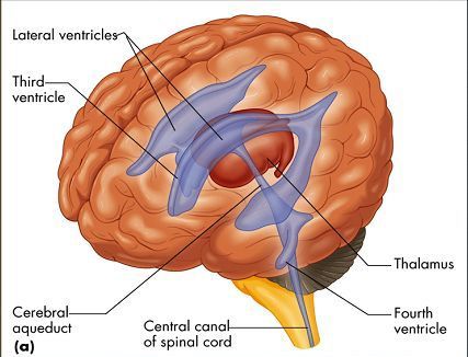
Fig 2.3 Ventricles of Brain.
The ventricular system comprises of –
a. Two
lateral ventricles
b. Single
3rd ventricle
c. Single
4th ventricle
All of these are connected to each other via aqueducts
and channels.
These channels are –
a. Interventricular
foramen of Monroe
b. Cerebral
aqueduct
c. Central
canal
All of these secrete CSF via the choroid plexus
present inside them which are nothing but a capillary network that filter the
blood and their ultrafiltrate is called as CSF. This CSF circulates through the
whole of Brain and spinal cord and is absorbed by small granulations present
called as arachnoid granulations.
These granulations can be seen in the figure 2.4

Fig 2.4 Arachnoid granulations.
Lateral Ventricle lies in the parietal lobe. Where
roof is mare by the corpus callosum, floor by thalamus, caudate nucleus, fornix
and choroid plexus, and the lateral wall by the narrow area meeting the floor
and roof.
There is a projection of the ventricle into the
frontal lobe called as anterior horn of the lateral ventricle, and a posterior
projection into the occipital lobe is called as the Posterior horn of the
lateral ventricle. There is another horn projecting into the temporal lobe of
the brain called as inferior horn.

Fig 2.5 Horns of Ventricle
The third ventricle is located in between the thalami,
which opens posteriorly into the 4th ventricle via the cerebral
aqueduct and the lateral ventricles open into the 3rd ventricle via
the foramen of Monroe. The 4th ventricle is located just behind the
brainstem which is projected into the cerebellum.
Brainstem consists of mid brain, pons and medulla
oblongata. These are located ventrally. Mid brain predominantly is made of the
cerebral peduncles (pedunculus Latin for footstalk).

Fig 2.6 Brainstem
The brainstem is most important because it contains
the centres for respiration, vomiting etc. Also, cranial nerves majority arise
from the brainstem. This also serves as a pathway for tracts to go up and down
the brain and periphery.
Mid brain has 4 bumpy structures in the posterior
aspect and are called as colliculi. They are 2 superior colliculi and 2
inferior colliculi. And we can study the brainstem at cross sectional levels at
these two colliculi.
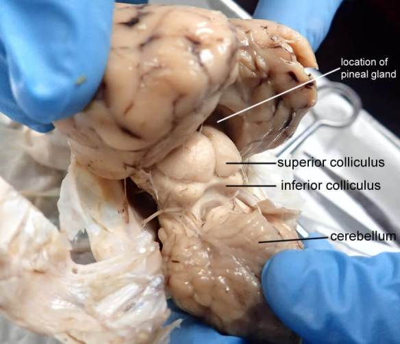
Fig 2.7 Colliculi of brain.
The blood supply to the brain is by the following
arteries –
a. Anterior
cerebral artery
b. Middle
cerebral artery
c. Posterior
cerebral artery
Anterior and Middle Cerebral arteries are a branch of
internal carotid arteries, whereas the posterior cerebral artery is a branch of
basilar artery which is intern an artery formed by the joining of the two
vertebral arteries.
This conjunction at the brainstem is called as circle
of Willis.

Fig 2.8 Circle of Willis.
This circle of Willis is an important aspect of
brain’s circulation because it ensures uninterrupted blood supply.
This circle is completed by the following arteries –
a. Anterior
communicating arteries
b. Posterior
communicating arteries
Venous drainage of brain is in the following order –
a. Veins
of brain
b. Intracranial
Dural sinuses
c. Internal
jugular veins of neck
d. Vena
cava
e. Right
atrium of heart
3 main veins run along the brain –
a. Superior
cerebral vein
b. Middle
cerebral vein
c. Inferior
cerebral vein
Other veins are –
a. Anterior
cerebral veins
b. Basal
vein of Rosenthal
c. Internal
cerebral veins
d. Great
cerebral vein
e. Basal
vein
f. Collicular
vein
g. Cerebellar
veins

Fig 2.9 Sinuses of brain

Fig 3.0 Veins of brain seen in an angiogram


Informative and precisely segregated sub topics, it's the need of hr.. keep up 🙌
ReplyDeleteNice bro
ReplyDeleteGazab bhai
ReplyDelete💯💯
ReplyDeleteGood initiative🤟
ReplyDeleteSahi hai💯keep going
ReplyDelete